Staying at the forefront of the optometric field is about providing the absolute best tools to help our patients live their best lives. At Central Coast Optometric Center, our experienced eye care professionals blend proven techniques with the latest technology to deliver affordable care for something undeniably priceless: your vision health.
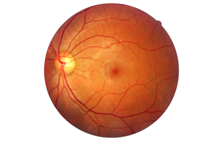
One of these important tools is fundus photography. A fundus camera is a specialized low power microscope with an attached camera designed to photograph the interior surface of the eye, including the retina, retinal vasculature, optic disc, macula, and posterior pole (i.e. the fundus).
Our optometrists use fundus photography to help get a deeper understanding of your ocular health. In addition to helping detect diseases early, retinal images provide a permanent and historical record of changes in your eye.
What are the benefits of Fundus Photography?
This picture allows your optometrist to see your eyes more closely and precisely. With the images produced, your doctor will be able to see early signs of eye diseases that they were unable to detect before, such as diabetic retinopathy, age related macular degeneration, macular edema and retinal detachment. Diagnosing conditions before they progress is important to avoid permanent side effects.
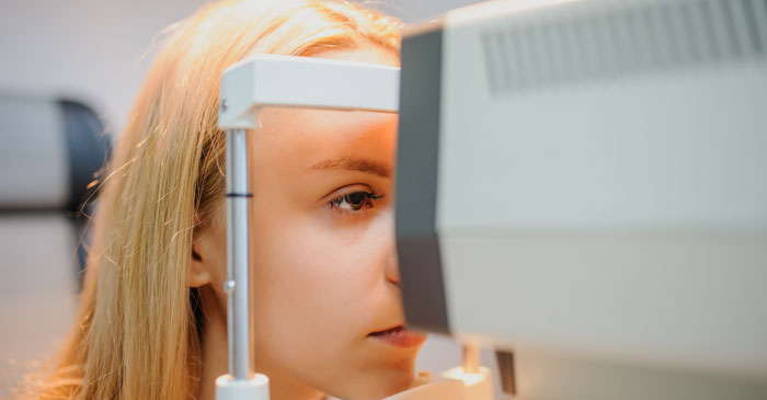
The fundus photography test is fast and painless, and results are seen almost immediately.
Fundus photos can be compared side-by-side over time to monitor your eye health and detect subtle changes. They allow your doctor to explain treatment more thoroughly as you can review the images together, which ensures a certain level of precision to your routine eye exam.
Who Needs Fundus Photography?
We strongly recommend that all of our patients receive a photograph of their eyes once per year. It is especially important for people who have:
- Headaches
- Spots or flashing lights
- Family or personal history of high blood pressure
- Circulatory problems
- Family or personal history of diabetes
- A strong eyeglass prescription
- Family or personal history of glaucoma
- History of choroidal nevus (freckle)