Blog
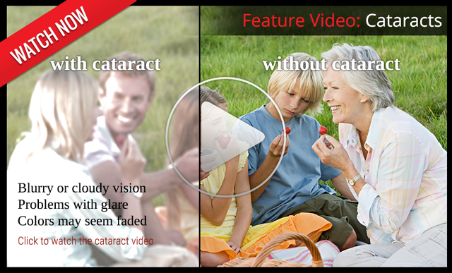
The Centers for Disease Control estimates that more than 2.8 million people in the United States suffer a concussion -- or traumatic brain injury (TBI) -- every year, and vision can be affected.
The rate of childhood TBI visits to the emergency department more than doubled between 2001 and 2009, making children more likely than any other group to go to the ER with concussion symptoms.
It was once assumed that the hallmark of a concussion was a loss of consciousness. More recent evidence, however, does not support that. In fact, the majority of people diagnosed with a concussion do not experience any loss of consciousness. The most common immediate symptoms are amnesia and confusion.
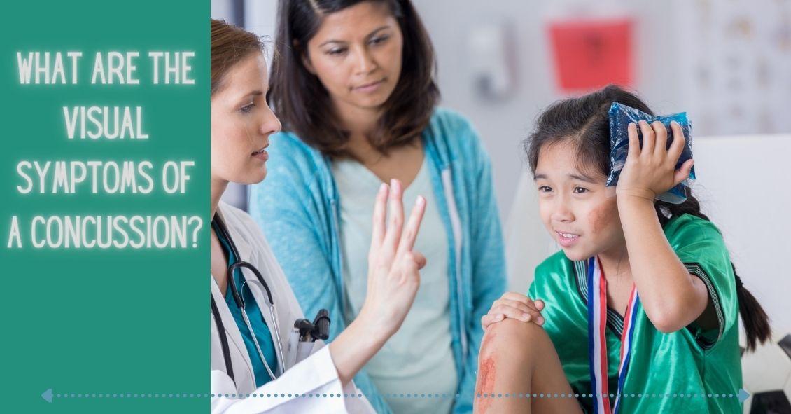
There also are multiple visual symptoms that can occur with a concussion, either initially or during the recovery phase.
Visual symptoms after a concussion include:
-
Blurred vision.
-
Difficulty reading.
-
Double vision.
-
Light sensitivity.
-
Headaches accompanying visual tasks.
-
Loss of peripheral vision.
Most people with visual complaints after a concussion have 20/20 distance visual acuity so more specific testing of near acuity, convergence amplitudes, ocular motility, and peripheral vision must be done.
In a study done at the Minds Matter Concussion Program at the Children's Hospital of Philadelphia, patients with a concussion diagnosis underwent extensive vision testing, which assessed symptoms, visual acuity, eye alignment, near the point of convergence, vergence amplitude and facility, accommodative amplitude and facility, and saccadic eye movement speed and accuracy.
A total of 72 children (mean age 14.6 years) were examined, and 49 (68%) of those were found to have one or more vision symptoms after concussion. The most common problems were convergence insufficiency (47.2%); accommodative insufficiency (33.3%); saccadic dysfunction (30.5%); and accommodative infacility (11.1%). The investigators also found that 64% of the children with convergence insufficiency also had an accommodative disorder.
Difficulties with accommodation and convergence make it very hard to read for any length of time, with blurring and fatigue and then loss of concentration occurring after a fairly short period of reading time.
For the majority of people suffering from a mild to moderate TBI, most of these symptoms resolve in one to three weeks but in some, they can persist much longer.
If your visual symptoms after a concussion persist past three weeks, a visit with an eye care specialist is recommended. There may be several options to help improve the symptoms with either prescription eyeglasses or prisms to assist the two eyes to focus together.
Article contributed by Dr. Brian Wnorowski, M.D.
This blog provides general information and discussion about eye health and related subjects. The words and other content provided in this blog, and in any linked materials, are not intended and should not be construed as medical advice. If the reader or any other person has a medical concern, he or she should consult with an appropriately licensed physician. The content of this blog cannot be reproduced or duplicated without the express written consent of Eye IQ.
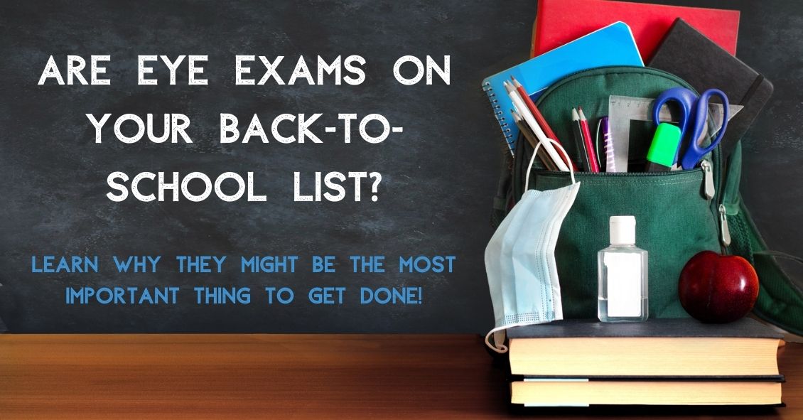
Is making an appointment for a comprehensive eye exam for your children on your back-to-school checklist? It needs to be.
No amount of new clothes, backpacks, or supplies will allow your child to reach their potential in school if they have an undetected vision problem.
The difference between eye exams and vision screenings
An annual exam done by an eye doctor is more focused than a visual screening done at school. School screenings are simply "pass-fail tests" that are often limited to measuring a child’s sight clarity and visual acuity up to a distance of 20 feet. But this can provide a false sense of security.
There are important differences between a screening and a comprehensive eye exam.
Where screening tests only for visual acuity, comprehensive exams will test for acuity, chronic diseases, color vision and eye tracking. This means a child may pass a vision screening at school because they are able to see the board, but they may not be able to see the words in the textbook in front of them.
Why back-to-school eye exams matter
Did you know that 1 out of 4 children has an undiagnosed vision problem because changes in their eyesight go unrecognized?
Myopia, or nearsightedness, is a common condition in children and often develops around the ages of 6 or 7. And nearsightedness can change very quickly, especially between the ages of 11 and 13, which means that an eye prescription can change rapidly over a short period of time. That’s why annual checkups are important.
Comprehensive eye exams can detect other eye conditions. Some children may have good distance vision but may struggle when reading up close. This is known as hyperopia or farsightedness. Other eye issues such as strabismus (misaligned eyes), astigmatism, or amblyopia (lazy eye) are also detectable.
Kids may not tell you they're having visions issues because they might not even realize it. They may simply think everyone sees the same way they do. Kids often give indirect clues, such as holding books or device screens close to their face, having problems recalling what they've read, or avoiding reading altogether. Other signs could include a short attention span, frequent headaches, seeing double, rubbing their eyes or tilting their head to the side.
What to expect at your child's eye exam
Before the exam, explain that eye exams aren’t scary, and can be fun. A kid-friendly eye exam is quick for your child. After we test how he or she sees colors and letters using charts with pictures, shapes, and patterns, we will give you our assessment of your child’s eyes.
If your child needs to wear glasses, we can even recommend frames and lenses that would be best for their needs.
Set your child up for success
Staying consistent with eye exams is important because it can help your kids see their best in the classroom and when playing sports. Better vision can also mean better confidence because they are able to see well.
Because learning is so visual, making an eye examination a priority every year is an important investment you can make in your child's education. You should also be aware that your health insurance might cover pediatric eye exams.
Set your child up for success and schedule an exam today!